
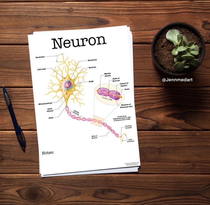
Delve into the intricate world of neurons with our comprehensive Anatomy of a Neuron Worksheet. This engaging resource unveils the secrets of these fundamental cells, providing a clear understanding of their structure, function, and role in the complex symphony of the nervous system.
Journey through the intricate workings of a neuron, unraveling the mysteries of dendrites, axons, and synapses. Discover the electrical properties that govern their communication and explore the fascinating realm of neurotransmitters, the chemical messengers that orchestrate intercellular dialogue.
Structure of a Neuron
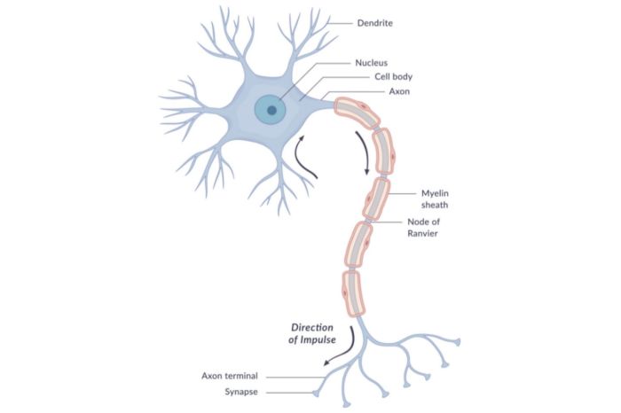
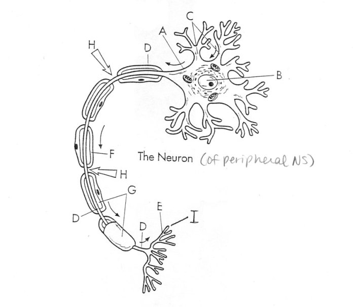
Neurons are the basic building blocks of the nervous system, responsible for transmitting information throughout the body. They consist of several distinct parts that work together to facilitate communication.
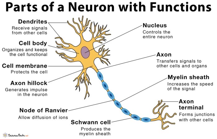
The main parts of a neuron include the cell body, dendrites, axon, axon hillock, myelin sheath, and axon terminals.
Cell Body
The cell body, also known as the soma, is the central part of the neuron. It contains the nucleus, which houses the cell’s genetic material, and other organelles essential for cell function.
Dendrites
Dendrites are short, branching extensions that extend from the cell body. They receive signals from other neurons and transmit them to the cell body.
Axon
The axon is a long, slender extension that carries signals away from the cell body. It is covered by a myelin sheath, which insulates the axon and speeds up signal transmission.
The anatomy of a neuron worksheet provides a thorough overview of the neuron’s structure and function. To test your understanding, check out the unit 1 progress check: frq . Completing the worksheet will reinforce your knowledge of the neuron’s components and how they contribute to the transmission of nerve impulses.
Axon Hillock
The axon hillock is the region where the axon originates from the cell body. It is where the electrical signals are generated that travel down the axon.
Myelin Sheath
The myelin sheath is a layer of fatty tissue that surrounds the axon. It acts as an insulator, increasing the speed of signal transmission.
Axon Terminals
Axon terminals are the ends of the axon that transmit signals to other neurons. They release neurotransmitters, which are chemical messengers that bind to receptors on the receiving neuron.
Types of Neurons

Neurons, the fundamental units of the nervous system, exhibit a remarkable diversity in structure and function. This diversity gives rise to the different types of neurons, each with specialized roles in processing and transmitting information.
Based on their structure, neurons can be classified into three main types: unipolar, bipolar, and multipolar.
Unipolar Neurons
- Unipolar neurons have a single process that extends from the cell body. This process functions as both the axon and dendrite, receiving and transmitting signals.
- They are primarily found in the embryonic nervous system and sensory ganglia of invertebrates.
Bipolar Neurons
- Bipolar neurons have two processes: a single dendrite and a single axon.
- They are found in the retina, cochlea, and olfactory epithelium, where they serve as sensory receptors.
Multipolar Neurons, Anatomy of a neuron worksheet
- Multipolar neurons have multiple dendrites and a single axon.
- They are the most common type of neuron and are found throughout the central and peripheral nervous systems.
- Multipolar neurons can be further classified based on the length of their axons:
- Golgi type I neurons: Have long axons that project to distant parts of the nervous system.
- Golgi type II neurons: Have short axons that connect to nearby neurons.
Electrical Properties of Neurons

Neurons exhibit unique electrical properties that enable them to transmit information efficiently. These properties involve the resting membrane potential and the generation of action potentials.
Resting Membrane Potential
When a neuron is at rest, its interior is negative relative to its exterior. This difference in electrical potential, known as the resting membrane potential, is maintained by the differential distribution of ions across the neuron’s membrane.
- Potassium ions (K+) are more concentrated inside the neuron than outside, while sodium ions (Na+) are more concentrated outside than inside.
- Leakage channels in the membrane allow K+ ions to diffuse out and Na+ ions to diffuse in, creating an imbalance of charges.
- The sodium-potassium pump actively transports 3 Na+ ions out of the neuron and 2 K+ ions into the neuron, maintaining the ion concentration gradients.
Action Potential Generation
When a neuron receives a stimulus that reaches a certain threshold, it triggers an action potential. This is a brief electrical impulse that travels along the neuron’s axon.
- The stimulus opens voltage-gated Na+ channels in the membrane, allowing Na+ ions to rush into the neuron.
- This influx of positive charges depolarizes the membrane, making the inside less negative.
- When the membrane reaches a threshold potential, voltage-gated K+ channels open, allowing K+ ions to flow out of the neuron.
- The efflux of positive charges repolarizes the membrane, making the inside negative again.
- The refractory period follows the action potential, during which the neuron is less excitable due to the inactivation of Na+ channels and the opening of K+ channels.
Neurotransmitters and Synaptic Transmission
At the heart of neuronal communication lies the intricate interplay between neurons and neurotransmitters. Neurotransmitters are chemical messengers that facilitate the transmission of signals across the synaptic cleft, the tiny gap between neurons.
Types of Neurotransmitters
Neurotransmitters can be broadly classified into two types based on their effects on the postsynaptic neuron:
- Excitatory neurotransmitters:These increase the likelihood of an action potential being generated in the postsynaptic neuron. Examples include glutamate, acetylcholine, and dopamine.
- Inhibitory neurotransmitters:These decrease the likelihood of an action potential being generated in the postsynaptic neuron. Examples include GABA (gamma-aminobutyric acid) and glycine.
Plasticity and Learning
The brain is not a static organ; it is constantly changing and adapting in response to experiences. This ability to change is known as plasticity, and it is essential for learning and memory. Synaptic plasticity is a specific type of plasticity that refers to changes in the strength of synapses, the connections between neurons.
These changes can be either long-term or short-term, and they are thought to underlie the formation and storage of memories.
Long-Term Potentiation
Long-term potentiation (LTP) is a long-lasting increase in the strength of a synapse. It is thought to be a cellular mechanism for learning and memory. LTP occurs when a synapse is repeatedly stimulated, causing a sustained increase in the release of neurotransmitters from the presynaptic neuron.
This increase in neurotransmitter release leads to a corresponding increase in the depolarization of the postsynaptic neuron, which in turn leads to an increase in the strength of the synapse.
Long-Term Depression
Long-term depression (LTD) is a long-lasting decrease in the strength of a synapse. It is thought to be a cellular mechanism for forgetting. LTD occurs when a synapse is repeatedly stimulated with a weak stimulus, causing a sustained decrease in the release of neurotransmitters from the presynaptic neuron.
This decrease in neurotransmitter release leads to a corresponding decrease in the depolarization of the postsynaptic neuron, which in turn leads to a decrease in the strength of the synapse.
Disorders of the Nervous System
The nervous system is a complex network of neurons that controls various bodily functions. However, several disorders can disrupt neuron function, leading to a range of neurological impairments.
Neurodegenerative Disorders
Neurodegenerative disorders are characterized by the progressive loss of neurons. Examples include:
- Alzheimer’s Disease:Affects memory, thinking, and behavior due to the accumulation of amyloid plaques and neurofibrillary tangles in the brain.
- Parkinson’s Disease:Results in movement problems, rigidity, and tremors due to the loss of dopamine-producing neurons in the substantia nigra.
- Multiple Sclerosis:An autoimmune disorder where the immune system attacks the myelin sheath surrounding neurons, disrupting nerve signals.
Traumatic Brain Injuries
Traumatic brain injuries (TBIs) occur due to a blow or jolt to the head, causing damage to neurons and surrounding tissues.
- Concussion:A mild TBI that can cause temporary loss of consciousness, headache, and confusion.
- Contusion:A more severe TBI involving bruising of the brain tissue, leading to swelling and damage.
- Diffuse Axonal Injury:A severe TBI where the axons of neurons are sheared, disrupting communication between neurons.
Treatment and Interventions
Treatment for neurological disorders depends on the specific condition and severity. Options may include:
- Medications:To manage symptoms, such as reducing inflammation or improving neurotransmitter function.
- Physical and Occupational Therapy:To improve mobility, balance, and cognitive function.
- Speech Therapy:To address communication difficulties.
- Surgical Interventions:In some cases, surgery may be necessary to remove tumors or relieve pressure on the nervous system.
FAQ Insights: Anatomy Of A Neuron Worksheet
What is the main function of a neuron?
Neurons are the fundamental units of the nervous system, responsible for transmitting information throughout the body. They receive, process, and transmit electrical and chemical signals, enabling communication between different parts of the body.
How do neurons communicate with each other?
Neurons communicate with each other through specialized junctions called synapses. At the synapse, neurons release chemical messengers known as neurotransmitters, which bind to receptors on the receiving neuron, triggering a response.
What are the different types of neurons?
There are various types of neurons, each with a distinct structure and function. Some common types include sensory neurons, motor neurons, and interneurons. Sensory neurons transmit sensory information from the body to the brain, motor neurons carry signals from the brain to muscles and glands, and interneurons facilitate communication within the brain and spinal cord.
